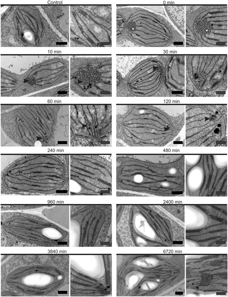
Figure 2.
Transmission electron micrographs of plastids present in chemically fixed deetiolating tobacco leaves. Etiolated leaf tissue contained plastids with structured, paracrystalline PLBs, in contrast with control plastids, harvested at the same time but not subjected to extended dark treatment. After just 10 min, the PLBs (stars) took on a less uniform, more irregular shape (arrowheads) and simultaneously began to disintegrate: residual PLBs were absent after 120 min of light. Subsequent time points were marked by increased abundance and size of grana membranes and the gradual accumulation of starch. Images at right reveal membrane structures at higher magnification. See Figure 3 and Supplemental Table S1 for quantification. For technical reasons (sample processing time), the 5 min time point was excluded from microscopy analysis. See Supplemental Figure S1 for comparison between chemical fixation and high-pressure freezing and Supplemental Figure S2 for further images of the etiolated plastids. Black bars = 500 nm and gray bars = 250 nm.

