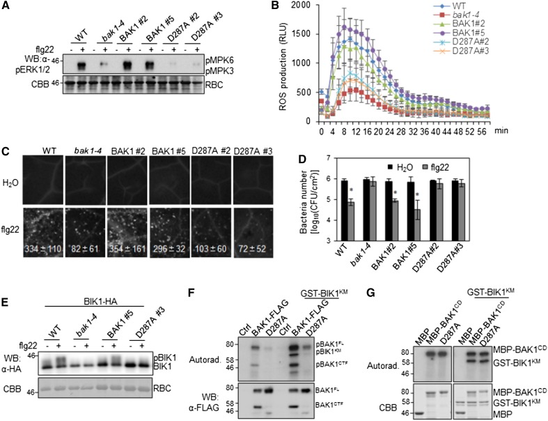
Figure 4.
Compromised immune responses in pBAK1::BAK1D287A/bak1 transgenic plants. A, flg22-induced MAPK activation. Two-week-old seedlings of wild-type Col-0, bak1-4, pBAK1::BAK1/bak1, and pBAK1::BAK1D287A/bak1 were treated with 100 nm of flg22 for 15 min. Phosphorylated MPK3 (pMPK3) and MPK6 (pMPK6) were detected by western blot (WB) with an α-pERK antibody (top). Protein loading is shown by Coomassie Brilliant Blue (CBB) staining for Rubisco (RBC; bottom). B, flg22-induced ROS production. Leaf discs from four leaves (technical repeats) of each of six 5-week-old plants (biological repeats) of indicated genotypes were treated with 100 nm of flg22, and ROS production was detected at the indicated time points. The data are shown as the mean ± sd from six biological repeats. C, flg22-induced callose deposition. Leaves of 4-week-old plants were collected for aniline blue staining 12 h after inoculation with 500 nm of flg22. Callose deposits were counted using the software ImageJ (1.43U). The data are shown as the mean ± sd from six biological repeats. D, flg22-mediated resistance to bacterial infection. Four-week-old plants were pretreated with 200 nm of flg22 or water and then infected with Pst DC3000 at 5 × 10⁵ cfu/mL. The bacterial growth assays were performed 2 d after infection. The data are shown as the mean ± sd from three biological repeats. The asterisks indicate statistical significance compared to H2O pretreatment by using Student’s t test (P < 0.05). E, flg22-induced BIK1 mobility shift. Arabidopsis protoplasts isolated from Col-0, bak1-4 mutant, pBAK1::BAK1/bak1, and pBAK1::BAK1D287A/bak1 transgenic plants were used to express BIK1-HA and treated with 100 nm of flg22 for 15 min. Mobility shift of BIK1 was detected by WB with an α-HA antibody (top). Equal loading of protein was indicated by CBB staining toward RBC (bottom). F, The kinase activity of BAK1 and BAK1D287A. FL BAK1-FLAG or BAK1D287A-FLAG were expressed in Arabidopsis Col-0 protoplasts and precipitated with an α-FLAG antibody. Kinase activity of the precipitated proteins were detected using GST-BIK1KM (kinase mutant) as a substrate. Phosphorylation was detected by autoradiography (top), and the protein loading is shown by WB with an α-FLAG antibody (bottom). G, The in vitro kinase activity of the cytosolic domain of BAK1 and BAK1D287A. GST-BIK1KM protein was used as a substrate and maltose-binding protein-BAK1CD (cytosolic domain) or its D287A mutant was used as the kinase in an in vitro kinase assay. Phosphorylation was detected by autoradiography (top), and the protein loading is shown by CBB staining (bottom). The above experiments were repeated three times with similar results. WT, wild type; pBIK1, phosphorylated BIK1.

