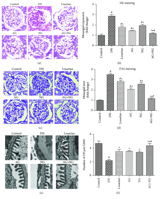
Figure 2.
Effects of AG and NG on renal histopathology and podocyte foot process effacement in diabetic rats. Representative hematoxylin and eosin (H&E, kidney histology (×400)) staining (a) and periodic acid-Schiff (PAS, kidney histology (×400)) staining (c) of kidney sections. Representative ultrastructure photos of glomerular podocytes taken by transmission electron microscopy (TEM) (×13500) (e). Semiquantitative analysis of mesangial expansion (b), mesangial area (d), and podocyte foot process density (f). FP: foot process; GBM: glomerular basement membrane; control: normal control rats; DN: STZ-induced diabetic rats; losartan: DN rats treated with losartan; AG: DN rats treated with Astragalus membranaceus; NG: DN rats treated with Panax notoginseng; AG+NG: DN rats treated with Astragalus membranaceus plus Panax notoginseng. Results were expressed as the mean ± SD. #P < 0.05 vs. control group, ∗P < 0.05 vs. DN group, ▲P < 0.05 vs. AG group, ▼P < 0.05 vs. NG group.

