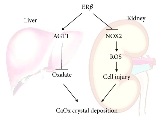
Figure 7.

Scheme. The schematic illustration of the ERβ signal regulation of CaOx crystal formation in the liver and kidney. For the liver part, ERβ signaling promotes activity of AGT1, which decreases the biosynthesis of oxalate by converting glyoxylate into glycine decreasing CaOx crystal formation. For the kidney part, ERβ has a protective effect in human renal tubular cells against oxalate-induced oxidative stress via suppression of NOX2 (a NADPH oxidase), which finally leads to decreasing CaOx crystal deposition on the damaged cell surface.
