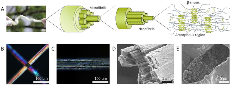
Figure 3. Hierarchical structure of A. yamamai silk fiber.

A, Schematic of the hierarchical structure of A. yamamai silk. B and C, The polarized light microscopy image of A. yamamai silk. D, Cross-sectional SEM image of A. yamamai silk after tensile fracture. E, Cross-sectional SEM image of A. yamamai silk fiber. The fiber was embedded in epoxy resin and further broken in liquid nitrogen.
