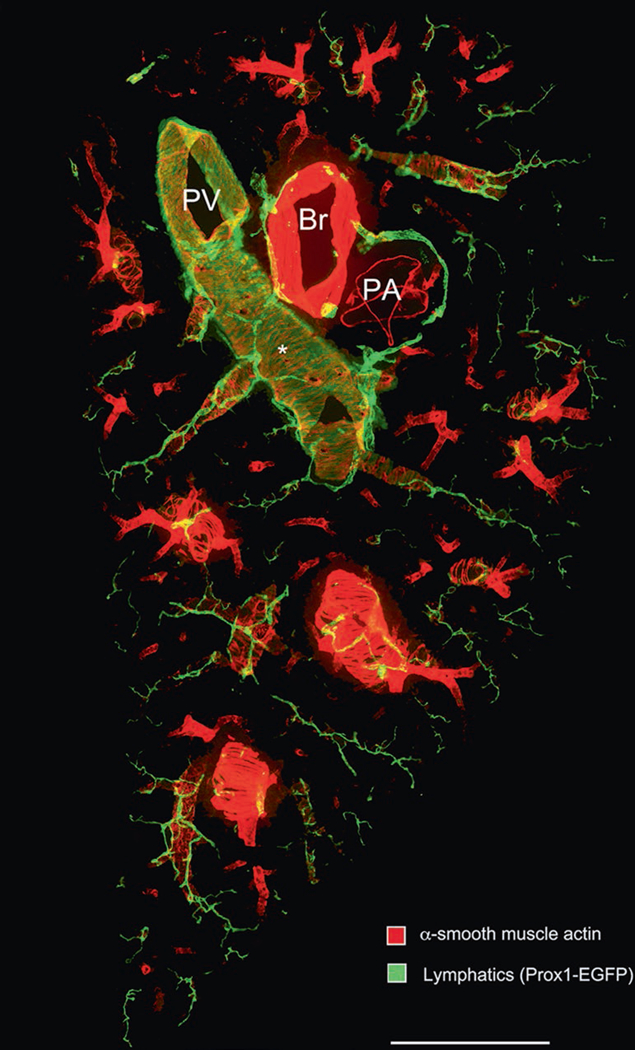
Fig. 2.

Low magnification panorama of 200-μm thick section of the entire width of the middle portion of left lung of a normal adult C57BL/6 Prox1-EGFP mouse prepared for immunohistochemistry according to the methods described. The section was stained for alpha-smooth muscle actin (red; Cy3- conjugated mouse anti-SMA, Sigma C6198) and Prox1-EGFP (green; Aves #1020 chicken anti-GFP). PV pulmonary vein, Br bronchus, PA pulmonary artery. (Asterisk) Cardiac muscle cells of the pulmonary vein express Prox1-EGFP, but are more weakly stained than the overlying lymphatics. Scale bar: 1 mm
