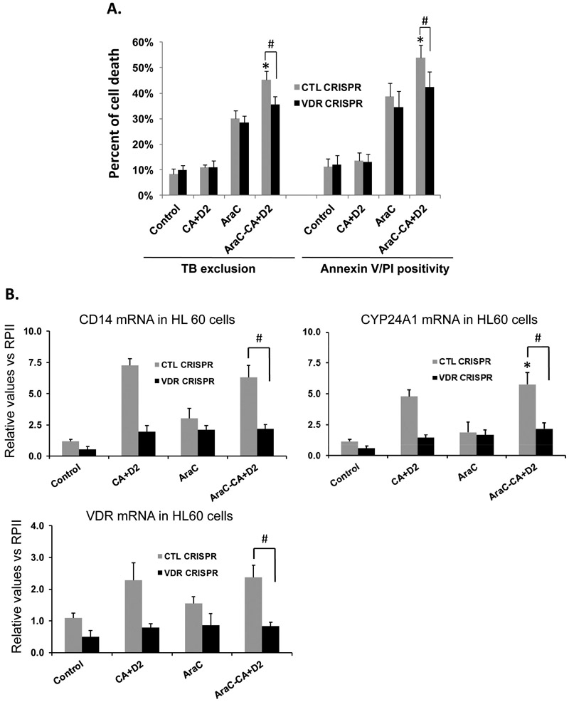
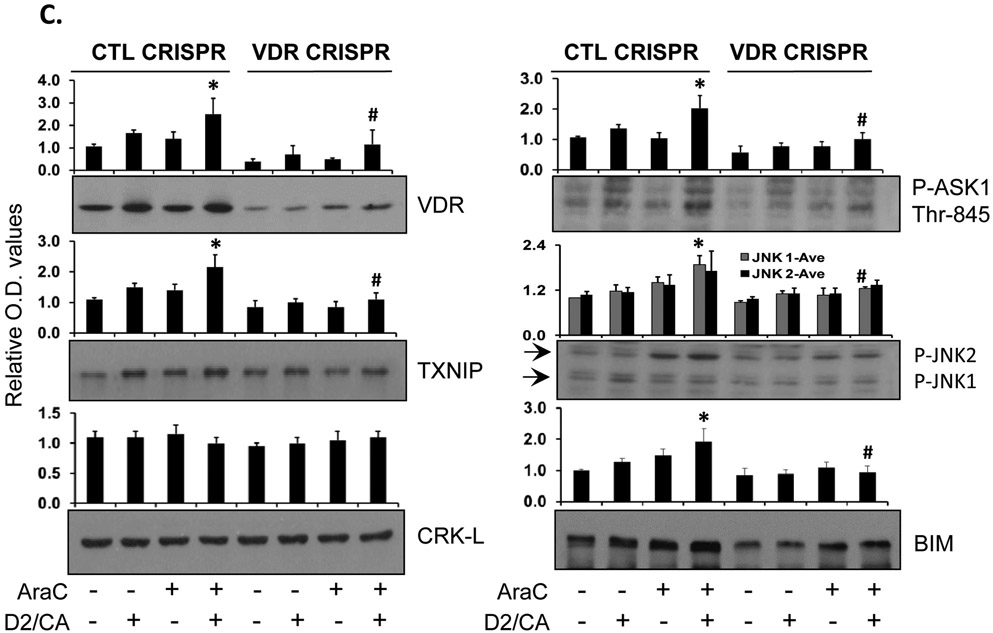
Figure 2. Knock down of VDR reduced cell death in the ECD phase in parallel with the reduction in apoptosis-related protein levels in HL60 cells.
A. VDR was knocked down by CRISPR/Cas9 in HL60 cells. Then the cells were pretreated with AraC (100 nM) for 72 h, followed by 100 nM 1-D2 and 10 μM CA (D2/CA) for 96 h. Percent of cell death determined by either Trypan blue exclusion or Annexin V/PI staining is shown in the bar chart. B. mRNA levels of VDR and its target genes CD14 and CYP24A1 in VDR KD cells (VDR CRISPR) as compared to the negative control cells (CTL CRISPR). C. A Western blot performed for the indicated apoptosis-related proteins. CRK-L was stripped and used as a loading control. The average relative signal intensities from three separate experiments are shown in bar charts above each blot. *, p<0.05 vs the AraC group; #, p<0.01 when comparing CTL CRISPR-ECD with VDR CRISPR-ECD.


