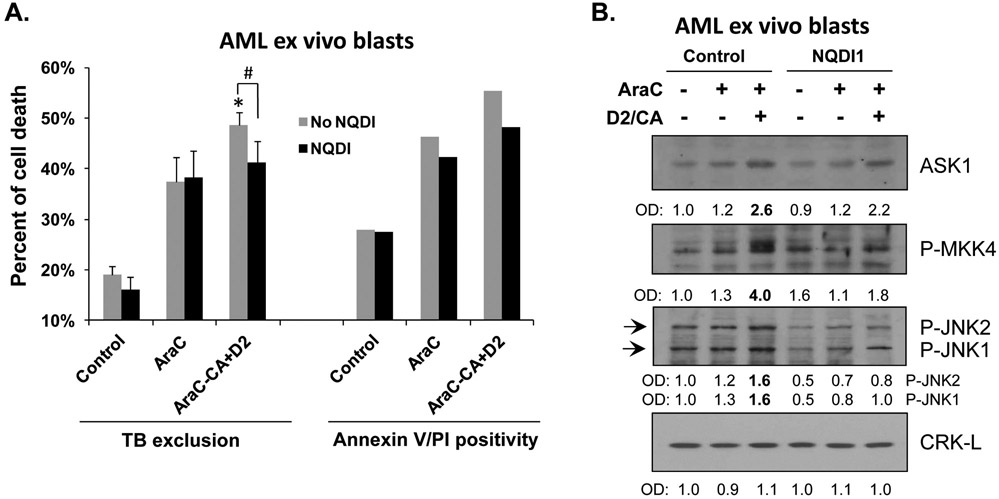
Figure 6. ASK1 contributes to ECD in AML ex vivo blasts.
A. Mononuclear cells were isolated from the bone marrow of a female AML-M4 patient. Cells were pretreated with either vehicle (Control) or 100 nM AraC, for 72 h. Both Control and AraC-treated cells were then post-treated for 96 h with ASK1 inhibitor NQDI-1 while D2/CA was added to a portion of AraC-treated cells during the post-treatment period, as indicted in the Figure. The percentages of dead cells determined by Trypan blue exclusion or annexin V/PI staining are shown in the bar chart. Since the ex vivo blast sample had limited size, we were not able to perform statistical analysis for Annexin V/PI assay. For Trypan blue exclusion assay, *, p<0.05 when compared to the AraC group; #, p<0.05 for the comparisons between the groups indicated in the chart. B. Western blots were performed for the indicated ASK1-related apoptosis signaling proteins. CRK-L was used as a loading control.

