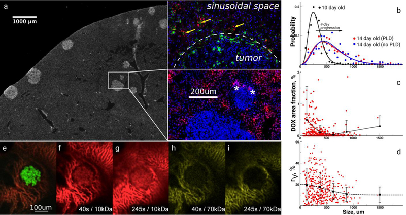
Figure 1. Heterogeneity of transport and structural properties of 4T1 breast cancer metastases in mouse liver.

a, Immunohistochemical image of a whole liver lobe scan containing metastases stained with 4’,6-diamidino-2-phenylindole. The inset shows several magnified metastases with different sizes and the red fluorescence of extravasated doxorubicin (DOX) delivered by PEGylated liposomal DOX (PLD) and colocalizing (yellow arrows) with Kupffer cells (green) outside tumors; stars denote DOX fluorescence in tumors, white-dashed line –tumor boundary. b, Size distribution of metastatic tumors in liver lobes before and after PLD as measured by imaging analysis. c, The area fraction of DOX inside tumors. Circles represent an individual tumor measurement followed by the average and standard deviation as a function of the sizes of metastases. d, The area fraction (%) of vasculature inside tumors stained with CD31. Circles represent an individual tumor measurement followed by the average and standard deviation as a function of the sizes of metastases. e, Intravital image (IVM) of the 4T1 tumor tagged with green fluorescent protein. IVM imaging of the extravasation of red 10 kDa (f, g) and yellow 70 kDa (h, i) dextran tracers.
