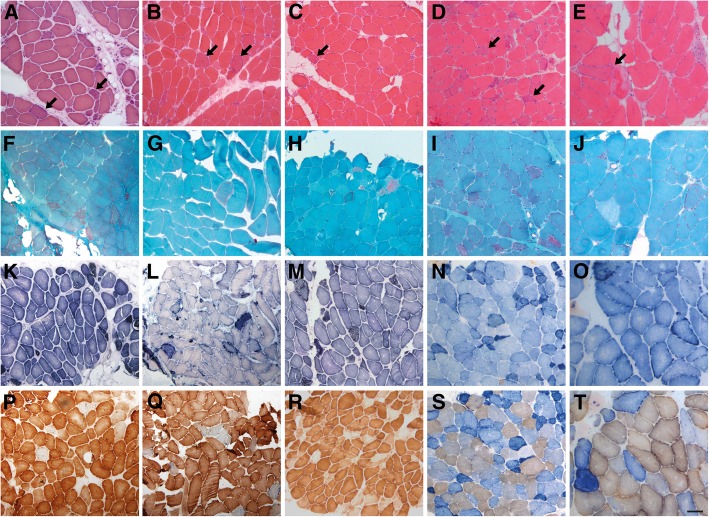
Fig. 2.
Morphological alterations in muscle biopsies from patients P1 (a, f, k, p), P5 (b, g, l, q), P9 (c, h, m, r), P14 (D, i, n, s) and P16 (e, j, o, t). a-e H&E shows dystrophic features in all cases with mild endomysal fibrosis, adipose tissue replacement, atrophy and necrotic fibers. Ragged-red fibers are frequently identified in all muscle samples (arrows). f-j Gomori trichrome showed the characteristic ragged-red fibers in all the biopsies. k-o Succinate dehydrogenase (SDH) reveals an increase of the oxidative staining in numerous fibers. p-t Frequent cytochrome C oxidase (COX) deficient fibers are present in variable proportion in the different cases (p and r, COX staining; o, s and t, COX-SDH combined staining). Scale bar =100 μm

