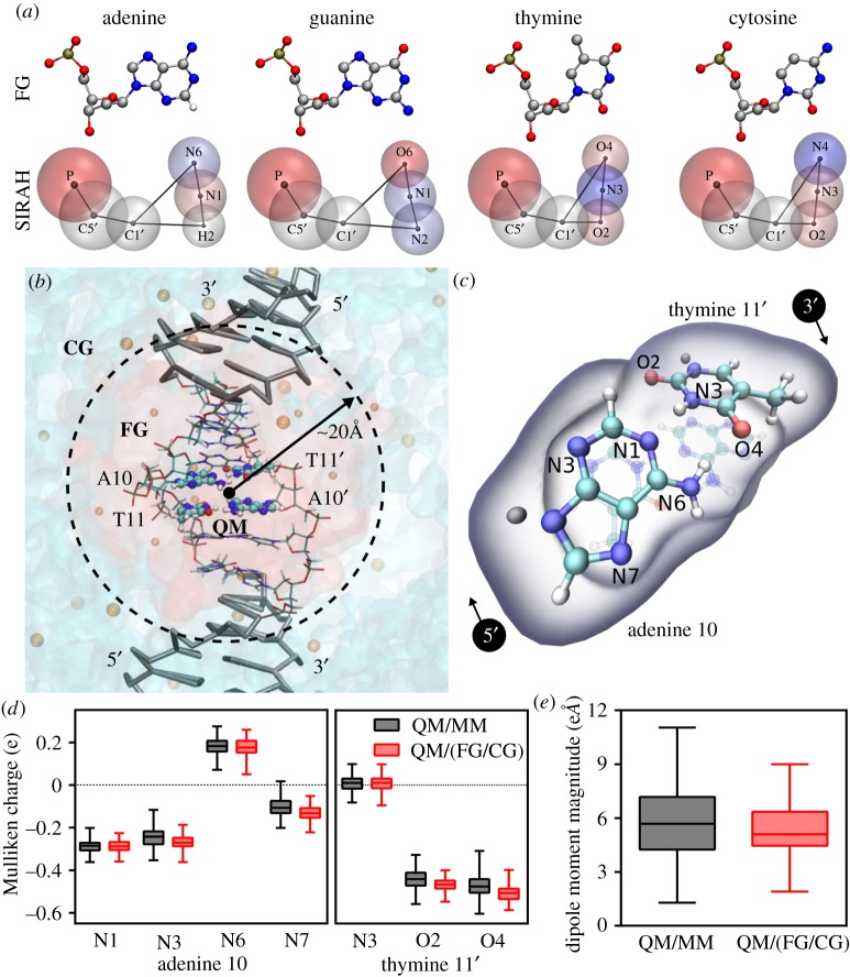
Figure 2.
Hybrid QM/(FG/CG) calculations in the 5′-CATGCATGCATGCATGCATG-3′ dsDNA. (a) DNA nucleotides at FG (top, only heavy atoms and hydrogen H2, which position is used for mapping in adenine are shown) and SIRAH representation (bottom, including mapped atom names). SIRAH beads are coloured according to their partial charge value, from negative (red) to neutral (white) and positive (blue). (b) Representation of the multi-scale partition of the system: QM treated atoms are shown as balls and sticks, FG base pairs are shown as thin sticks coloured by atom and CG DNA is represented as thick grey sticks. Transparent red and light blue surfaces represent the volume occupied by FG and CG solvent, respectively. FG and CG ions are shown as orange balls with different sizes. Owing to limited mixing of FG and CG solvents just an indicative FG/CG a dashed circle denotes interface. (c) Insight into the QM subsystem indicating the atom nomenclature. Link atoms are depicted in grey. (d) Comparison of Mulliken charges from 2000 single point calculations at QM/MM and QM/(FG/CG) level. (e) Comparison of the calculated dipole moment magnitude at the entire QM subsystems. (Online version in colour.)

