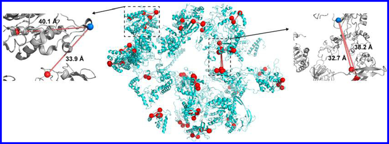
Figure 4.
Structure of DSSO cross-linked E. coli ribosome (PDB: 3jcd) with cross-linked lysine residues and distances shown. Cross-links are identified using MetaMorpheusXL at a 1% FDR cutoff. The whole E. coli ribosome structure is shown in the middle; red lines indicate Cα−Cα distances between the cross-linked lysine (marked as red spheres). Four cross-link outliers are also shown: at left panel with an inter-cross-link with Cα−Cα distance of 40.1 Å on P0A7V0(36) and P0A7V0(58) and an intra-cross-link with Cα−Cα distance of 33.9 Å on P0A7V0(36) and P0A7W7(69); at right panel with an intra-cross-link with Cα−Cα distance of 38.2 Å on P0A7L0(141) and P0A7N9(58) and another intra-cross-link with Cα−Cα distance of 32.7 Å on P0A7L0(141) and P0A7N9(9).

