Fig. 3.
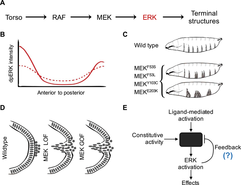
Quantitative studies in the early embryo allow identification of divergent effects of pathogenic mutations. (A) Simplified schematic of Torso RTK signaling in the early Drosophila embryo. (B) Schematic representation of the spatial profile of active, dually phosphorylated ERK (dpERK) for wild-type (solid) and pathogenic MEK variants (dotted) in the early embryo. (C) Schematics representing the larval cuticle phenotype for wild type and various gain-of-function (GOF) MEK mutants. (D) Pole-hole phenotype as a readout of the pathway function for MEK loss-of-function (LOF) (via MEK RNAi), and MEK GOF (via GOF mutations F53S, F53L, Y130C, and E203K). (E) Proposed negative-feedback based model to explain the divergent effects of activating mutations on ERK activation

