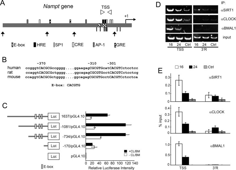
Figure 3. Regulation of the Nampt promoter by CLOCK:BMAL1 and SIRT1.
(A) Schematic diagram of regulatory elements in human Nampt promoter. TSS (Transcription Start Site) primer region for q-PCR is shown as arrowheads. Truncated forms of Nampt promoter used in (C) are shown (arrows). Putative transcription factors binding sites are indicated: HRE, hypoxia-inducible factor-responsible element; SP1, specificity protein 1; CRE, cAMP-response element; AP-1, activator protein 1; GRE, glucocorticoid receptor response element. (B) Conserved E-boxes (capitalized) are shown. Numbers indicate positions from human transcription start site. (C) Schematic diagram of Nampt promoter constructs. Effect of CLOCK:BMAL1 (+CL/BM; black bars) on luciferase activity is shown. Luciferase activity of CLOCK:BMAL1 on the pGL4.10 was set as 1. All data are means ± SEM of three independent samples. (D) Representative results of ChIP assays analyzed by semiquantitative PCR. Dual crosslinked nuclear extracts isolated from MEFs after 16 or 24 hr serum shock and subjected to ChIP assay with anti-SIRT1, anti-CLOCK, anti-BMAL1, or no antibody (ctrl). No antibody and 3’R primers were used as controls for immunoprecipitation and PCR, respectively. (E) Quantification of ChIP by q-PCR. q-PCR was performed on the same samples as described in (D). All data are means ± SEM of three independent samples.

