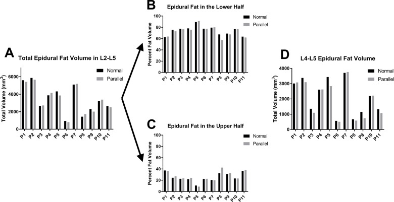
Figure 2.
Comparison of epidural fat quantification among MRI techniques. (A) Graphical comparison of epidural fat between L2 and L5 among the 11 patients using both the conventional MRI (normal) and parallel axial slices showed no significant difference in the fat volume calculations. (B, C) The epidural fat volume was split between L3 and L4 to illustrate the higher portion of fat in the lower section of the spine. (D) The distribution of L4–L5 epidural fat volume, shown here, was specifically compared because that is the location where the two MRI techniques differ the most due to the slant in the conventional protocol.

