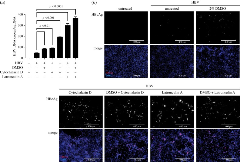
Figure 4.
Disruption of actin filaments increases the susceptibility of HepG2–NTCP cells to HBV infection. (a) HBV DNA secretion and (b) HBcAg immunofluorescence staining of untreated HepG2–NTCP cells or cells cultured in the presence of 2% DMSO, and/or 4 µg ml−1 Cytochalasin D or 0.1 µg ml−1 Latrunculin A, for 24 h following infection using 1000 GE/cell HBV. HBV DNA and HBcAg were determined 5 days post infection of cells. Data shown are mean (s.d.) of three independent experiments.

