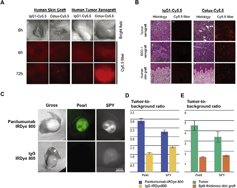
Fig. 3. Antibody-dye conjugates in tumor imaging.

(A) Stereomicroscopic imaging of engrafted human skin xenografts and human tumor explant xenografts in SCID mice injected with cetuximab conjugated with Cy5.5 (Cetux-Cy5.5) or nonspecific human IgG1 antibody (IgG1-Cy5.5). (B) Head-to-head comparison of the histology and fluorescent results in tumor sections. (C) Uptake of panitumumab-IRDye800 versus non-specific IgG-IRDye 800 using the Pearl and SPY Systems in mice with flank SCC1 tumors. (D) The tumor-to-background ratio in mice injected with panitumumab-IRDye800 or IgG-IRDye800. (E) Panitumumab-IRDye800 demonstrated significantly higher fluorescent signal in the tumor than the split-thickness skin graft in both the Pearl and SPY imager. Adapted from Refs. [75 and 77] with permissions.
