Figure 2.

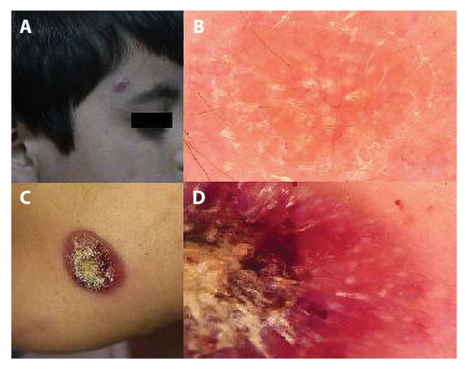
(A,C) Clinical picture of CL lesion. (B) Dermoscopically lesion reveals yellow tears (PlCD × 30). (D) Yellow tears with central crust (PlCD × 30). [Copyright: ©2019 Serarslan et al.]

(A,C) Clinical picture of CL lesion. (B) Dermoscopically lesion reveals yellow tears (PlCD × 30). (D) Yellow tears with central crust (PlCD × 30). [Copyright: ©2019 Serarslan et al.]