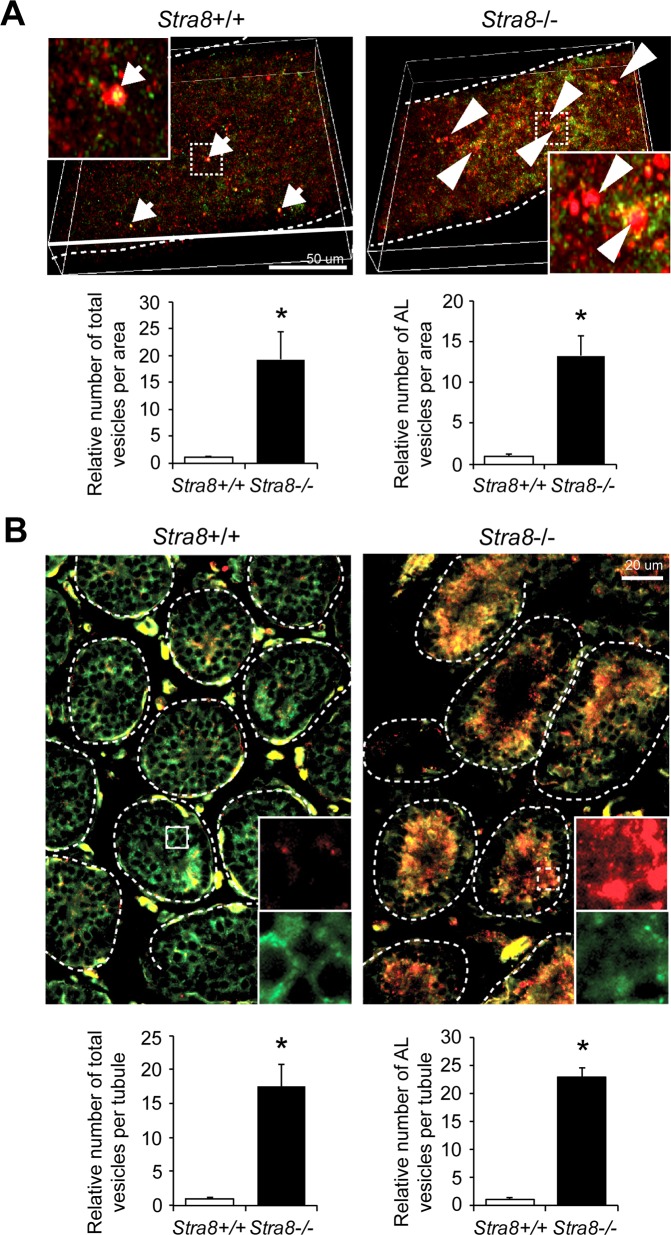
Fig 2. In vivo RFP-GFP-LC3 reporter in wild-type and Stra8-deficient testes.
(A) Whole-mount seminiferous tubules from RFP-GFP-LC3 transgenic mouse testes in juvenile wild-type and Stra8-deficient backgrounds by confocal imaging (upper panels). Quantification of vesicle numbers per imaging area relative to wild-type samples is shown. Total, RFP-positive vesicles. AL (Autolysosome), RFP-positive GFP-negative vesicles. All values are means ± SD. n = 3 mice per genotype. *P < 0.05 (Student’s t test). (B) Testicular cross sections of RFP-GFP-LC3 transgenic mouse testes in juvenile wild-type and Stra8-deficient backgrounds. Dashed lines indicate seminiferous tubules. Arrows indicate autophagosome. Arrowheads indicates autolysosome (autophagosome maturation). Quantification of vesicle numbers per imaging area relative to wild-type samples is shown. Total, RFP-positive vesicles. AL (Autolysosomes), RFP-positive GFP-negative vesicles. All values are means ± SD. n = 3 mice per genotype. *P < 0.05 (Student’s t test).

