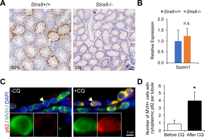
Fig 3. Rapid autophagic degradation of p62 in Stra8-deficient testicular germ cells.
(A) Immunohistochemistry of p62 in wild-type and Stra8-deficient testes at 21 d.p.p.. Numbers indicate the percentages of seminiferous tubules containing germ cells with nuclear p62 accumulation. (B) qRT-PCR analysis of Sqstm1 in wild-type and Stra8-deficient testes normalized to β-actin. Data represent mean ± SD; n = 3 per group. n.s.: not significant. (C) Dual immunofluorescence staining of p62 and MVH in Stra8-deficient testes treated with vehicle (PBS; left panel) or chloroquine (CQ; right panel) treatment for 7 days. (D) Quantification of the number of MVH+ germ cells exhibiting cytoplasmic p62 expression per seminiferous tubule with or without chloroquine treatment in Stra8-deficient testes. Data represent mean ± SD; n = 3 animals per group; 326 seminiferous tubules were examined from Stra8-deficient testes without chloroquine treatment. 296 seminiferous tubules were examined from Stra8-deficient testes with chloroquine treatment. *P < 0.05 (Student’s t test).

