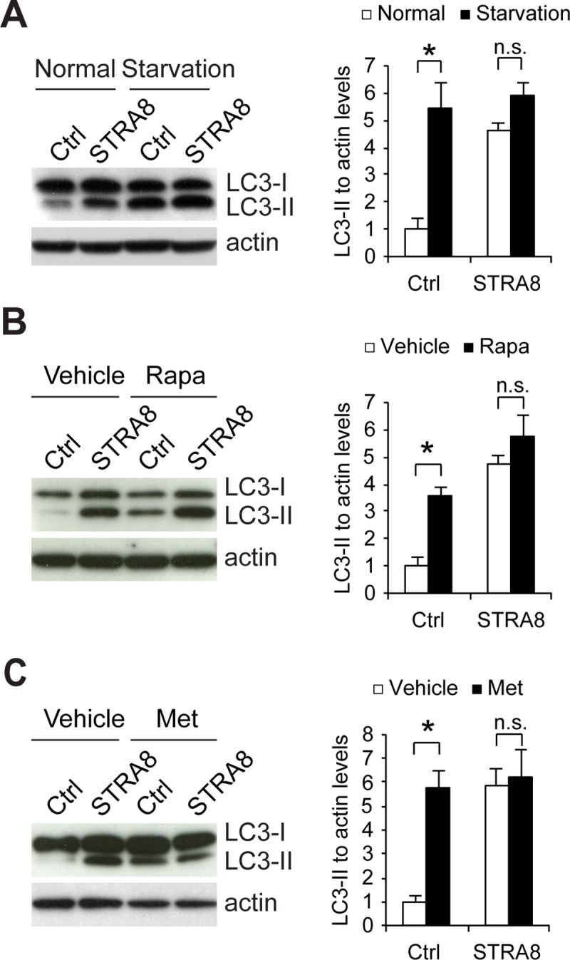
Fig 5. STRA8 inhibits de novo autophagosome formation upon autophagy induction.

(A) Cell lysates from F9 cells stably expressing GFP (Ctrl) or STRA8 (tagged with GFP) treated with EBSS for 2 hours were subjected to Western blot analyses using antibodies as indicated. Graph shows quantification of LC3-II to actin ratio. Data represent mean ± s.e.m; n = 3 independent experiments; *P < 0.05 (Student’s t test). (B) Cell lysates from F9 cells stably expressing GFP (Ctrl) or STRA8 (tagged with GFP) treated with vehicle or rapamycin (Rapa; 0.1 μM) for 2 hours were subjected to Western blot analyses using antibodies as indicated. Graph shows quantification of LC3-II to actin ratio. Data represent mean ± s.e.m; n = 3 independent experiments; *P < 0.05 (Student’s t test). (C) Cell lysates from F9 cells stably expressing GFP (Ctrl) or STRA8 (tagged with GFP) treated with vehicle or metformin (Met; 2 mM) for 2 hours were subjected to Western blot analyses using antibodies as indicated. Graph shows quantification of LC3-II to actin ratio. Data represent mean ± s.e.m; n = 3 independent experiments; *P < 0.05 (Student’s t test).
