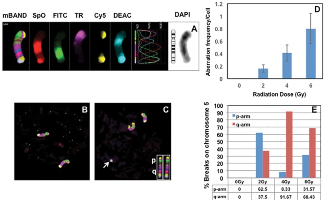
Fig 5. Detection of intra-chromosomal aberrations in G0 PCCs using chromosome specific mBAND probe.

(A) The hybridization pattern of five different fluorochromes [SpO- Spectrum Orange, FITC-Fluorescein isothiocyanate, TR-Texas Red, Cy5- Cyanine 5 and DEAC-7-diethylaminocoumarin; DAPI (4′, 6-diamidino-2-phenylindole-chromosome counterstain]. Representative G0 PCCs of control (B) and irradiated (C; 4 Gy of γ-rays) lymphocytes probed with chromosome 5 specific mBAND probe are shown. Note the terminal fragment of one of the chromosomes 5 (arrow) in the irradiated G0 PCC spread. The hybridization patterns observed in the p- and q-arms of metaphase chromosome 5 are shown in the insert. (D) Frequency of total intra-chromosomal aberrations (fragments of p and q arms, translocations and inversions) detected for different γ-rays doses in G0 PCCs. (E) Frequency of breaks observed in the p- and q-arms of the chromosome 5 detected by the mBAND technique. The percentage of breaks observed in the short and long arms of chromosome 5 for different γ-rays doses is shown in the form of histogram. Bars represent SEM.
