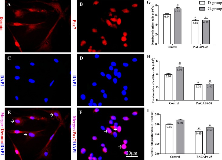
Fig 5. Effect of PACAP6-38 on number and proliferation of satellite cells in pectoral muscle.
Identification of satellite cells by immunofluorescent staining of desmin (A: Desmin; C: DAPI; E: Merger) and Pax7 (B: Pax7; D: DAPI; F: Merger) in primary cultured cells. (G) Relative number of satellite cells. (H) Absolute number of satellite cells. (I) Proliferation of satellite cells. Data are mean ± SEM. # p < 0.05 Vs. D-group control; * p < 0.05 Vs. different reagent treatment in the same light groups. Desmin labeling the cellular structure (A, red), Pax7 expressed in cell nucleus (B, red), and nuclei are stained with DAPI (C and D, blue). The arrows indicate the positive cells (E, F). Green light increased the relative numbers, absolute numbers and proliferation of satellite cells compared with D-group. PACAP6-38 decreased the relative numbers, absolute numbers and proliferation of satellite cells, and the differences of these disappeared between D-group and G-group. Scale bar = 20 μm.

