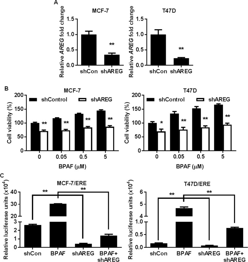
Fig 7. AREG knockdown impedes BPAF-mediated cell proliferation and ER transcriptional activity.
MCF-7 and T47D cells were transiently transfected with lentivirus-mediated AREG shRNA. PCR validation of AREG knockdown is shown in A. B) Control and AREG shRNA MCF-7 and T47D cells were serum-starved for 48 hours in phenol red-free medium. Then, cells were treated with BPAF (0, 0.05, 0.5, or 5 μM) in phenol red-free medium with 5% C.S. FBS for 5 days. The average percentage of viable cells in each treatment group was determined with an MTT assay. C) Control and AREG shRNA MCF-7 and T47D cells were transiently transfected with the ERE luciferase reporter plasmids, followed by serum starvation in phenol red-free medium for 48 hours. Then, the cells were exposed to BPAF (1 μM) in phenol red-free medium with 5% C.S. FBS for 24 hours. The relative luciferase activities for each treatment group are graphed. All values are presented as the means ± S.E. (**P<0.01).

