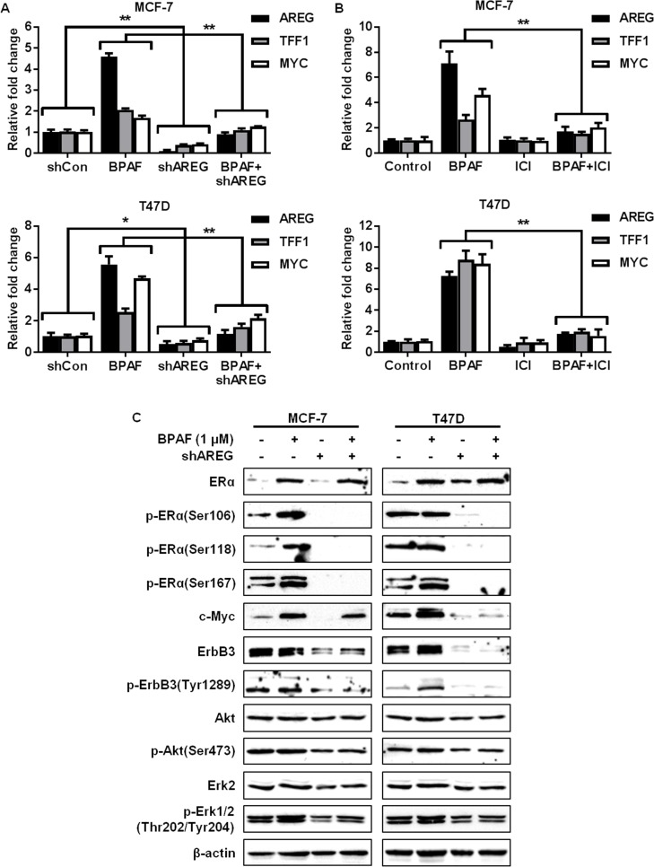
Fig 8. AREG knockdown blocks BPAF-mediated ER-RTK crosstalk.
Control and AREG shRNA MCF-7 and T47D cells were serum-starved for 48 hours in phenol red-free medium. Then, cells were treated with BPAF (0 or 1 μM) in phenol red-free medium with 5% C.S. FBS for 16 hours (A) and MCF-7 and T47D cells were treated with BPAF (1 μM) ± ICI-182,780 (2 μM) in phenol red-free medium with 5% C.S. FBS for 16 hours (B), followed by qPCR analysis of AREG, TFF1, and MYC, the ER/RTK target genes most significantly upregulated by BPAF. The fold changes for the treated samples are graphed relative to the normalized values of the corresponding controls. Values are presented as the means ± S.E. (*P<0.05, **P<0.01 as compared to the corresponding treatment groups). C) Control and AREG shRNA MCF-7 and T47D cells were serum-starved for 48 hours in phenol red-free medium. Then, cells were treated with BPAF (0 or 1 μM) in phenol red-free medium with 5% C.S. FBS for 30 minutes. Then, Western blotting analysis was performed on the indicated markers involved in the ER and ErbB3/RTK signaling pathways.

