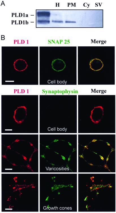
Figure 1.
PLD1 localization to the neuronal plasma membrane. (A) Brain synaptosomes lysed by hypotonic shock were homogenized (H) and processed to separate the cytosol (Cy), the membrane-bound compartment (PM), and the crude synaptic vesicle fraction (SV). Proteins (10 μg) were subjected to gel electrophoresis and immunodetection on nitrocellulose sheets by using anti-PLD1 Abs. Lysates from HEK293 cells transfected with pCGN-PLD1a and pCGN-PLD1b were used as positive controls. (B) Immunofluorescent confocal micrographs of cultured cerebellar granule cells double-labeled with anti-PLD1 Abs (red) and anti-SNAP-25 or anti-synaptophysin Abs (green). (Bar = 5 μm.)

