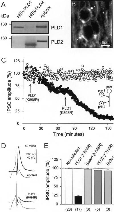
Figure 2.
Injection of catalytically inactive PLD1 inhibits evoked-ACh release in Aplysia neurons. (A) Lyzed Aplysia ganglia membrane-bound proteins (20 μg) were subjected to gel electrophoresis and immunodetection by using anti-PLD1 or anti-PLD2 Abs. Lysates from HEK293 cells expressing PLD1 or PLD2 were used as controls. (B) Immunofluorescent confocal micrographs of Aplysia buccal ganglion neuropile labeled with anti-PLD1 Abs. Note that PLD1 outlined axon (arrows). (C) Amplitude of IPSCs evoked at identified synapses in the buccal ganglion of Aplysia was averaged for periods of 10 min (mean ± SD) and plotted against time. PLD1(K898R) was pressure injected (arrow) in one of the presynaptic neurons (●). The second presynaptic neuron was not injected (○). (D) Representative presynaptic action potentials and IPSCs recorded before (control) and 120 min after PLD1(K898R) injection. (E) ACh release measured 120 min after injection and expressed relative to the initial response obtained before injection. For control, neurons were injected with PLD2(K758R), buffer, or boiled PLD1(K898R). Values in brackets indicate the number of experiments.

