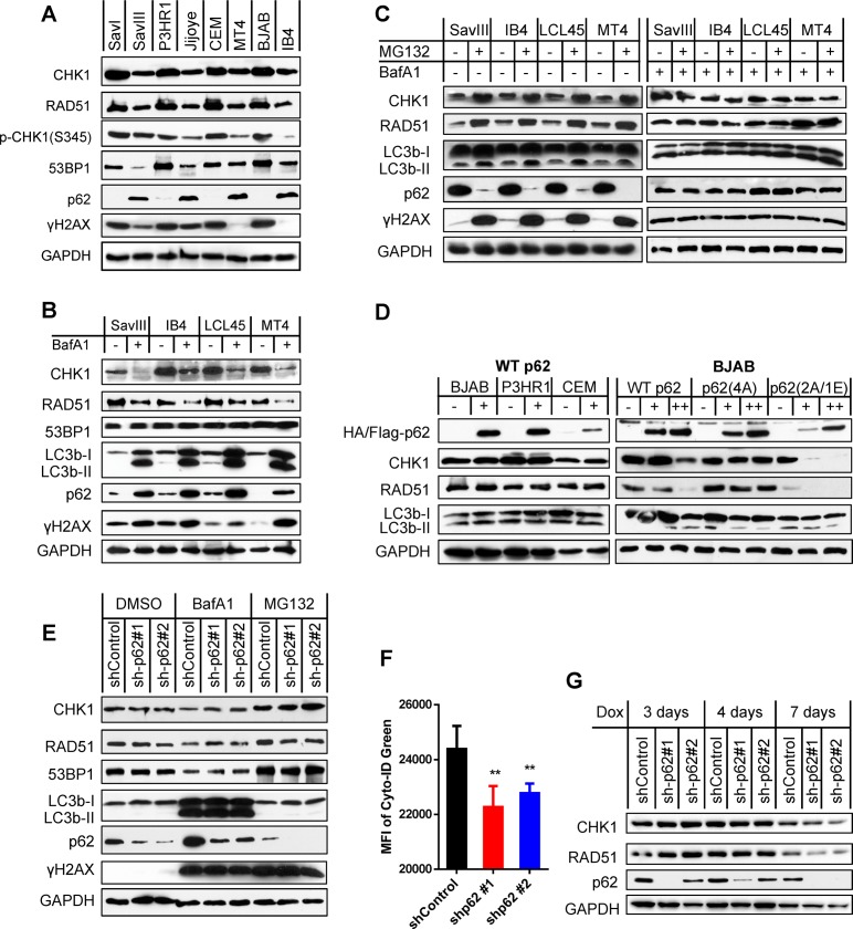
Fig 5. p62 accumulation upon autophagy inhibition destabilizes HR DNA repair proteins CHK1 and RAD51 in viral latency.
A. Correlation of p62 with CHK1, RAD51 and 53BP1 protein levels in viral latency were analyzed in paired cell lines. B. Cell lines with higher levels of p62 protein were treated with 0.4 µM BafA1 (Sigma) for 48 h. Indicated proteins were probed by immunoblotting. C. Cell lines with higher levels of p62 protein were treated with 10 µM of the proteasome inhibitor MG132 for 6 h (left panel), or pre-treated with 0.4 µM BafA1 before MG132. Indicated proteins were probed by immunoblotting. D. Cell lines with lower levels of p62 protein were transfected with 5 µg (+) or 10 µg (++) of HA-p62 plasmids, its mutants with Flag tag, or vector control in each electroporation (1X10⁷ cells). Indicated proteins were analyzed by immunoblotting 48 h post-transfection. E-F. IB4 cells stably harboring p62 shRNA or shRNA control in 1 µg/ml puromycin were treated with 0.4 µM BafA1 for 48 h or 10 µM MG132 for 6 h, before subjected to immunoblotting or flow cytometry. shRNA expression was induced by 1 µg/ml doxycycline for 2 days before the drug treatments. MFI = mean fluorescence intensity. G. CHK1 and RAD51 protein stability was evaluated by immunoblotting in virus-transformed IB4 cells stably expressing control shRNA or p62 shRNA that were induced by 1 µg/ml doxycycline for different time points.

