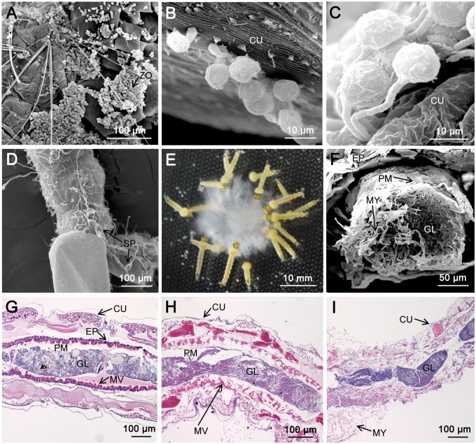
Fig 1. Two infection pathways by P. guiyangense into mosquito larvae.
(A-D) Infection by direct penetration of the mosquito cuticle. (A) Zoospores gathered and adhered to the cuticle of a larva at 4 hpi. (B) Germinating cysts with germ tubes and appressoria at 8 hpi. (C) Germ tubes penetrating larvae at 12 hpi. (D) Sporangia produced at 24 hpi. (E-I) Infection by disruption of the mosquito digestive tract. (E) Mosquito larvae devouring P. guiyangense mycelia. (F) The mosquito larvae midgut packed with mycelia at 2 hpi. (G-H) The mosquito midgut epithelium, muscles, and connective tissues appeared disrupted at 12 hpi and 24 hpi. (I) Complete disruption of the mosquito epithelium and peritrophic membrane after 48 h. ZO: zoospore, MY: mycelia, SP: sporangia, CU: cuticle, EP: epithelium, PM: peritrophic membrane, GL: gut lumen, MV: microvilli.

