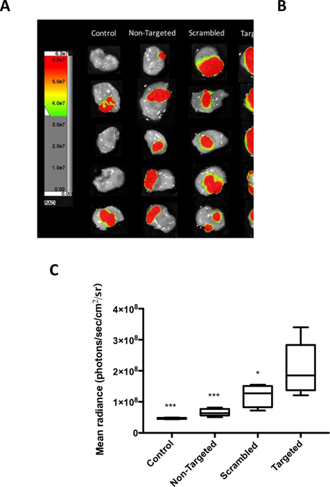
Figure 7.
In vivo bladder tumor tissue targeting evaluation using double PEGylated liposomes incorporating different functionalized lipopolymers (non-targeted, 2 mol% mPEG2000-DSPE; scrambled, 2 mol% WVRF-PEG2000-DSPE; or targeted, 2 mol% RWFV-PEG2000-DSPE) versus untreated controls. A: Luciferin-induced luminescence signal from MB49-Luc bladder tumor cells; B: Cy5.5 fluorescence arising from liposomes bound to bladder tissue; and C: Mean radiance (whole bladder ROI’s) of control and treated tissues using different liposome preparations. The liposomes were instilled into the bladder intravesically and allowed to in-dwell for 1 h before aspirating the liposome suspension and washing the bladder with PBS. Luciferin was then injected i.p. before euthanasia. The bladder was then harvested, inverted, washed, and placed for imaging. Liposome diameter: 61 nm. Liposome formulation: 58.5:35:4:2:0.5 DPPC:Chol:mPEG1000-DSPE:X:Cy5.5-DSPE, where X = mPEG2000-DSPE (non-targeted); WVRF-PEG2000-DSPE (scrambled); or RWFV-PEG2000-DSPE (targeted). Control: no liposome treatment. Error bars indicate the standard deviation of the mean with n=5 (ANOVA: *p < 0.05, **p < 0.01, ***p < 0.005, ****p < 0.001).

