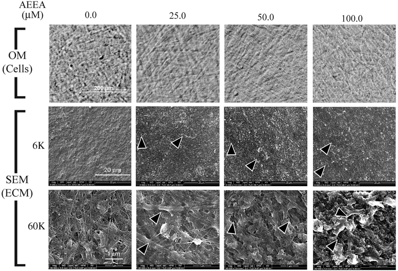
Figure 2. AEEA markedly altered the structure of the extracellular matrix produced in vitro.
Upper panel: Cells treated with AEEA (0.0, 25.0, 50.0, or 100 μM) for 14 days were observed under an optical microscope (OM) before decellularization. Middle and lower panels: ECM was examined with scanning electron microscopy (SEM) under low power (× 6,000 magnifications, middle panel) or high power (× 60,000 magnifications, lower panel). (See Results for detailed description).

