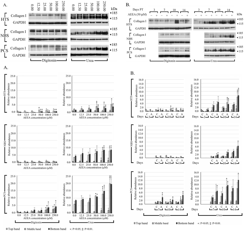
Figure 4. Effects of AEEA on extractability of type I collagen, and collagen deposition in three human fibroblast cell lines.
(A) Cells were treated with varying concentrations of AEEA for 10 days. (B) Time course of the effects of AEEA on the amount of extractable type I collagen protein. Cells were treated with 50 μM AEEA and harvested at the indicated time. Digitonin: samples extracted with native lysis buffer containing 1% digitonin. Urea: samples extracted with urea lysis buffer after native lysis buffer extraction. Densitometry analysis: relative abundances were normalized to the top band of the control sample of the digitonin fraction of each cell line, respectively. These results were representative of at least three independent biological replicates.

