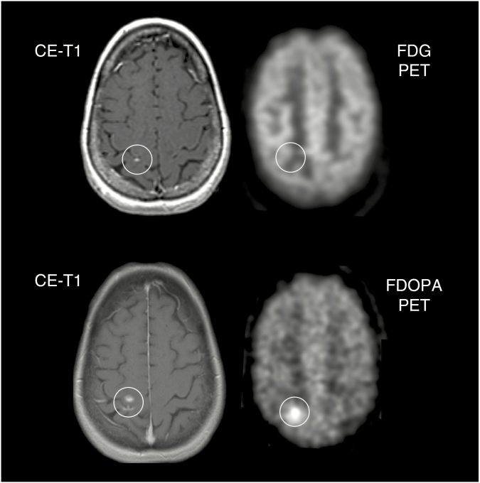
Fig. 1.
A 56-year-old female patient with a brain metastasis originating from a papillary thyroid carcinoma treated with radiosurgery. Follow-up MR imaging 15 months later (top row, left) is consistent with stable disease according to RANO criteria for brain metastases. Most probably due to the lesion size, the corresponding FDG PET (top row, right) shows no increased metabolic activity. During the next 12 months, the size of contrast enhancement increased marginally (bottom row, left). Notwithstanding the small lesion size on anatomical MRI, the corresponding FDOPA PET (bottom row, right) shows clearly increased metabolic activity indicating brain metastasis relapse.

