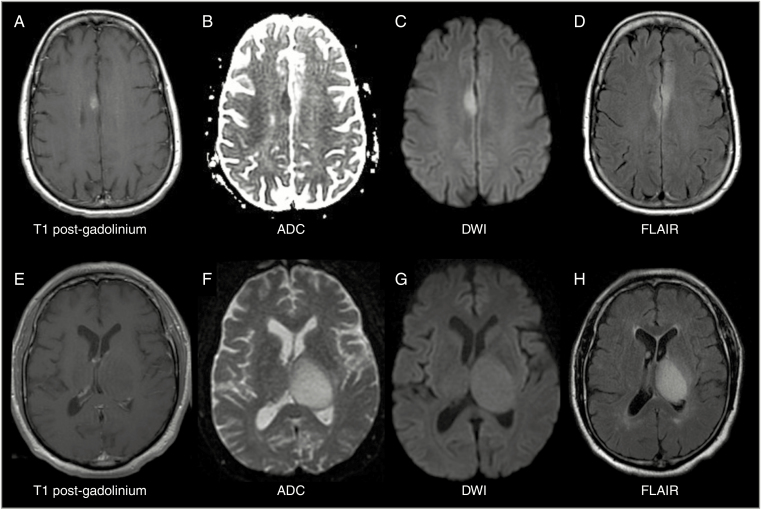
Fig. 2.
Representative spectrum of MRI neuroimaging features in EGFR-amplified, IDH-wildtype lower-grade glioma. Top row (A–D) shows example of a T1-weighted post-contrast enhancing (A), diffusion restricted (B, C), FLAIR infiltrative (D), EGFR-amplified anaplastic astrocytoma. Bottom row (E–H) shows an example of a non-enhancing (E), diffusion facilitated (F, G), and expansile (H) EGFR-amplified anaplastic astrocytoma. Abbreviations: ADC, apparent diffusion coefficient; DWI, diffusion-weighted image; FLAIR, fluid-attenuated inversion recovery.

