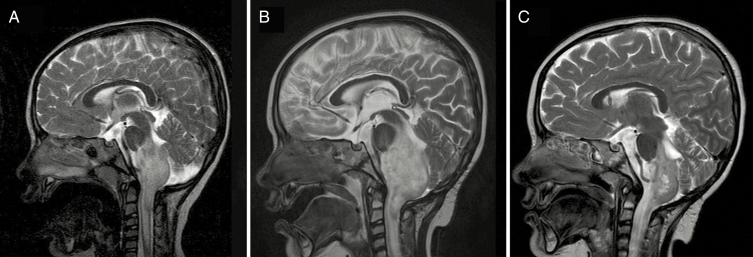
Fig. 2.
Case of severe PsP toxicity. A 3.9-year-old female patient presenting with cervicomedullary WHO grade II fibrillary astrocytoma treated with 50.4 Gy(RBE) PBT with subsequent symptomatic PsP requiring ventricular shunt and permanent tracheostomy. T2 mid-sagittal MRI images shown. (A) Pre-PBT baseline scan demonstrates infiltrative cervicomedullary tumor. (B) Four-month post-PBT scan demonstrates increased size of infiltrating tumor (ventricular shunt placed prior to this scan). (C) Twelve-month post-PBT scan demonstrates spontaneous decreased size of tumor; ventricular shunt remains in place.

