Figure 2.
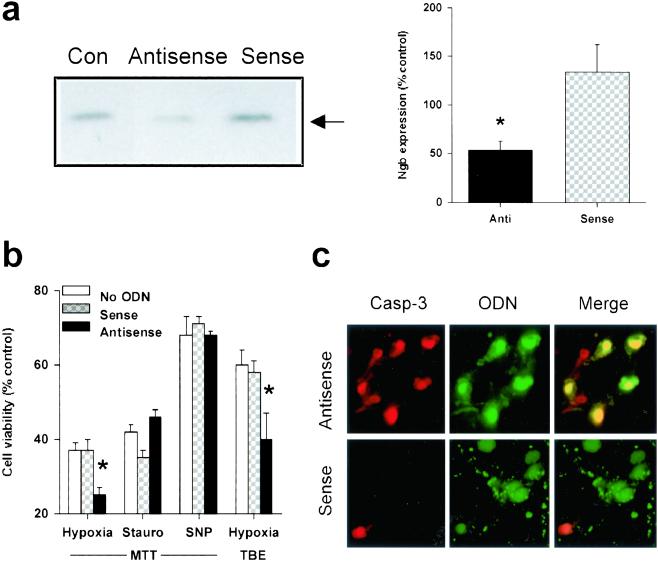
Decreased Ngb expression exacerbates hypoxic neuronal death. (a) Representative Western blot showing decreased Ngb expression compared to untransfected control cells (Con) in cultured cortical neurons treated with an antisense ODN directed against Ngb, but not with a sense ODN (Left). Ngb expression was quantified (mean ± SEM, n = 3) by computer densitometry (*, P < 0.05 relative to Con by t test) (Right). (b) Cell viability, measured by MTT absorbance or TBE, in cultures maintained for 12 h without oxygen or in the presence of 0.1 μM staurosporine (Stauro) or 200–400 μM SNP, under standard conditions (no ODN) or after treatment with 5 μM sense or antisense ODN, added 3 h before the onset of, and present throughout the toxic exposure (n = 3–6). *, P < 0.05 relative to no treatment (t test). (c) Fluorescence labeling of cultured cortical neurons treated with Ngb antisense (Upper) or sense (Lower) ODNs, showing immunoreactivity for the 17–20-kDa caspase-3 cleavage product (Left, red), ODN fluorescence (Center, green), and the merged images (Right, yellow). (Original magnification, ×400). Antisense-transfected cultures show an increase in the proportion of neurons that exhibit caspase-3 cleavage compared to sense-transfected cultures, consistent with the antisense-mediated decrease in cell viability shown in b.

