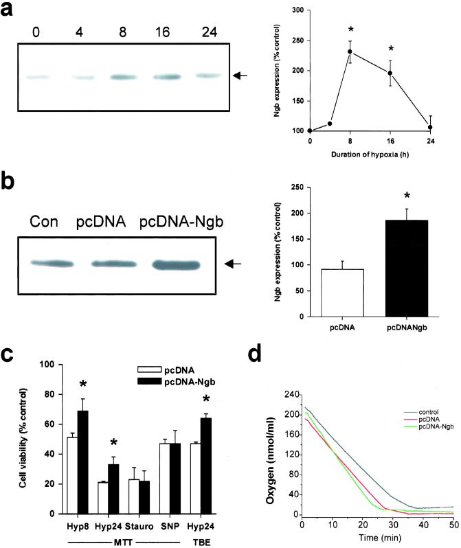
Figure 3.
Overexpression of Ngb reduces hypoxic cell death. (a) Representative Western blot showing increased Ngb expression in cultured HN33 cells maintained without oxygen for the indicated number of hours (Left). Expression of the 17-kDa band (arrow) was quantified by computer densitometry (mean ± SEM, n = 3; *, P < 0.05 relative to 0 h by t test) (Right). (b) pcDNA vector or Ngb-expressing recombinant plasmid (pcDNA-Ngb) was stably transfected into HN33 cells, and the overexpression of Ngb protein in pcDNA-Ngb-transfected cultures was confirmed by Western blotting (Left). Ngb expression was quantified by computer densitometry (mean ± SEM, n = 5; *, P < 0.05 relative to Con by t test) (Right). (c) Cell viability, measured by MTT absorbance or TBE, in pcDNA- or pcDNA-Ngb-transfected cultures maintained for 8 h (Hyp8) or 24 h (Hyp24) without oxygen, or for 24 h in the presence of 0.1 μM staurosporine (Stauro) or 300 μM SNP. *, P < 0.05 relative to untransfected control cultures (t test). (d) Oxygen consumption in untransfected (control) and pcDNA- or pcDNA-Ngb-transfected HN33 cells (5 × 10⁶ cells in 1 ml of DMEM) measured by using a Clark oxygen electrode (Hansatech Instruments, Pentney King's Lynn, U.K.). Each tracing is the average of three independent experiments. The slopes give oxygen consumption (nmol O/ml/min) and were not significantly different across conditions (control, 7.46 ± 0.90; pcDNA, 9.24 ± 1.39; pcDNA-Ngb, 11.87 ± 2.31; P = 0.18 by ANOVA, n = 9).

