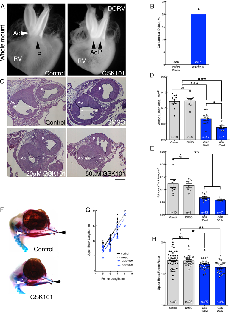
Fig. 4. Ligand activation of TRPV4 replicates hyperthermia induced birth defects.

(A) Whole mounts of hearts from control and GSK101 treated-embryos. GSK-treated heart showed DORV orientation of the aorta (Ao) and pulmonary trunk (P). See fig. S9 for histological sections. Whole mount and histological analysis of heart anatomy was performed in 38 hearts from DMSO-treated embryos and 15 GSK101-treated embryos.(B) Percentage of hearts with histologically confirmed conotruncal defects in GSK101 (GSK)-treated embryos compared to DMSO controls. (C) Panel of histological sections through the aorta (Ao) and pulmonary trunk (P) comparing the luminal areas (white arrows) at the level of the left coronary artery (*) in control, DMSO, and 20μM or 50μM GSK101 treatment. Bar = 200μm. (D,E) Treatment with GSK101 reduced aortic luminal areas (D, average coefficient of error (CE)=0.03) and pulmonary trunk luminal areas (E, average CE=0.03). (F) Alcian blue and alizarin red stains of DMSO control (top) and 20μM GSK101 treated embryos (bottom) at HH36. (G) Normalization of upper beak length to femur lengths. (H) Graph of upper beak to femur ratios in untreated control, DMSO-treated control and 2 GSK101-treatment groups. Significance was determined using Fisher exact test (B) or one-way ANOVA followed by Bonferroni’s multiple comparisons test (D, E, H). *P<0.03, **P<0.004, ***P<0.0001. The number of biological replicates is indicated by n in the graphs in (D), (E), and (H).
