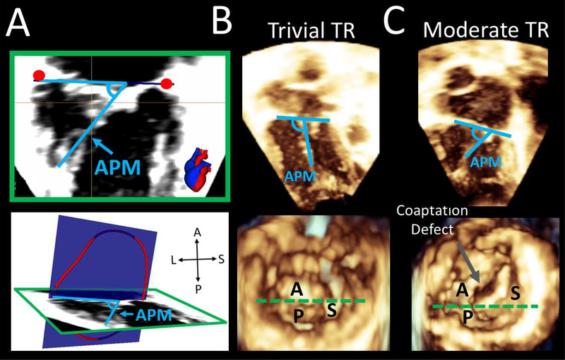
Figure 5. Measurement and Comparison of Anterior Papillary Muscle Angles During Systole.
A. Measurement of anterior papillary muscle (APM) angle relative to annular plane in apical 4 chamber view and ventricular 3D view; B. APM measurement from 4 chamber and ventricular 3D view in a patient with trivial TR. Note obtuse APM angle relative to annular plane; C. 4 chamber and ventricular view in patient with moderate TR. Note acute APM angle relative to annular plane and coaptation defect. Dotted green line in Figures B and C represents plane in Figure A ventricular view. A= anterior leaflet; P = posterior leaflet; S = septal leaflet; APM = anterior papillary muscle; TR = tricuspid regurgitation.

