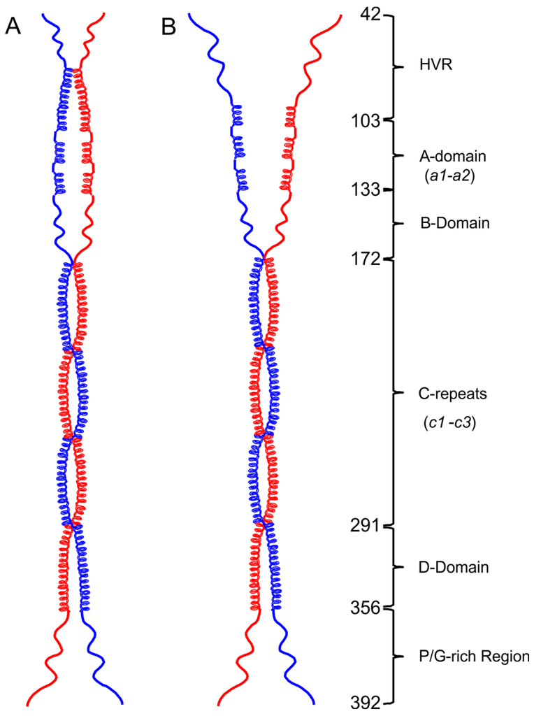
Fig. 6. Open and closed conformations of the PAM structural model.
Dimeric models were drawn to scale in ChemDraw Professional 16.0 based upon the domain organization of PAMAP53. (A) At 4° C, the HVR with some α-helix content dimerizes and forms a closed pattern at the NH2-terminus. (B) At 25° C or 37° C, loss of α-helices within the HVR regions occur, eventually resulting in an open status of NH2-terminal domains, viz., HVR-A-B domains. The numbers of the first residue in each domain are listed on the illustration.

