
Figure 1. DPSC morphology during expansion.
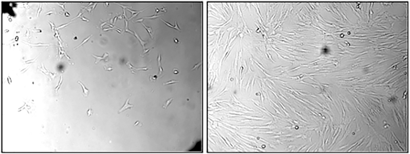
Dental pulp tissues were plated to expand stem cells. Left panel shows early growth of stem cells and right panel shows stem cells after confluence.
Official websites use .gov
A
.gov website belongs to an official
government organization in the United States.
Secure .gov websites use HTTPS
A lock (
) or https:// means you've safely
connected to the .gov website. Share sensitive
information only on official, secure websites.
Dental pulp tissues were plated to expand stem cells. Left panel shows early growth of stem cells and right panel shows stem cells after confluence.