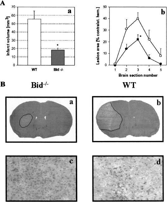
Figure 7.
(A) Ischemic damage is reduced in Bid−/− mice. (a) Infarct volume was reduced by 67% in Bid−/− mice as compared with wt mice (n = 8 per group; P < 0.01) after 30 min of MCAo and 48 h of reperfusion. (b) Protection in Bid−/− mice was significant at the level of the striatum (section 3; P < 0.01) with a trend of infarct size difference at other brain section levels. (B) Ischemic tissue damage in Bid−/− and Bid+/+ mice. As shown by representative H&E-stained coronal-brain sections, Bid−/− mice had significantly smaller infarcts after 30 min of MCAO and 48 h of reperfusion (a) as compared with Bid mice (b). High-power micrographs (×200) taken adjacent to penumbral areas (cortical layer III) showing little evidence of cellular damage in Bid−/− mice (c), and extensive injury in wt mice (d).

