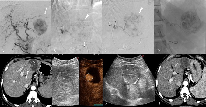
Fig. 3.
A 156-year-old male with a single nodule of HCC with maximal diameter of 76 mm at II/III hepatic segments. Figure A, Digital subtraction angiography (DSA) performed from the common hepatic artery, shows the hypervascular structure of the HCC in the left lobe (arrow). Super-selective DSA of the tumour with deflated balloon figure B) and inflated balloon figure C) (arrowhead). Figure D shows single fluoroscopy image after the embolization. Figure E, 1-month follow-up CT in arterial phase, highlights the partial necrosis of the nodule (star) with the presence of a hypervascular bottom (arrow) of vital residual tumour. Figure F, contrast-enhanced ultra-sound, shows the residual HCC, and figure G evidences the radiofrequency ablation of the lesion (arrow). Figure H, post-procedural CT in arterial phase, evidences the complete response of the HCC

