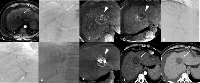
Fig. 4.
A 42-year-old female with HCC at level of the IV hepatic segment (maximal diameter 42 mm). Figure A, magnetic resonance imaging, arterial phase, shows non-homogeneous hypervascular nodule in the IV segment (arrow). The tumour is confirmed by the digital subtracted angiography (DSA) (figure B, arrow), in the cone-beam CT arterial phase (figure C) (arrowhead) and cone-beam CT delayed phase (figure D) (arrowhead). Super-selective DSA of the tumour with deflated balloon figure E) and inflated balloon (arrow) figure F). Figure G shows single fluoroscopy image after the embolization. Figure H non-enhanced cone-beam CT at the end of the procedure shows complete filling of the HCC (qTCR) (arrowhead). 1-month follow-up CT in arterial phase (figure I) and late phase (figure L) demonstrates the complete response of the HCC (arrow)

