Fig. 4.
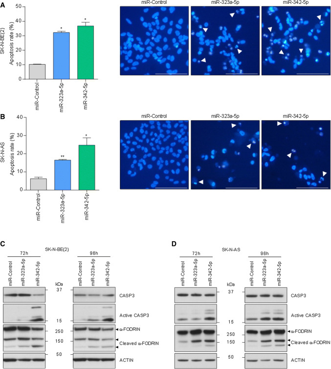
MiR-323a-5p and miR-342-5p overexpression induced apoptosis in NB cells. a, b Analysis of chromatin fragmentation/condensation in SK-N-BE(2) (a) and SK-N-AS (b) transfected with 25 nM of miR-control, miR-323a-5p or miR-342-5p 96 h post-transfection. Images on the right show a representative field of NB cells stained with Hoechst dye. White arrowheads point to cells with condensed and/or fragmented chromatin. Data represent mean ± SEM of three independent experiments (n = 3 per experiment). *p < 0.05, **p < 0.01, two-tailed Student’s t test. c, d Representative Western blot analysis of apoptosis-related proteins in SK-N-BE(2) (c) and SK-N-AS (d) transfected with miR-control, miR-323a-5p or miR-342-5p (25 nM) at 72 h and 96 h post-transfection

