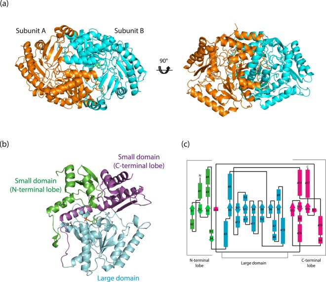
Figure 1.
Overall structure of Tr-ωTA. (a) Overall structure of Tr-ωTA. The structure as a dimer form is depicted as a cartoon and viewed from two different directions. Subunits A and B are coloured orange and cyan, respectively. The PMP molecule in subunit B is represented as green sticks. (b) The two domains of Tr-ωTA (subunit B): large domain (residues 94–315; cyan), small domain (residues 1–93 and 316–451). The small domain is divided into two lobes (N- and C-terminal lobes): N-terminal lobe (residues 1–93; green) and C-terminal lobe (residues 316–451; magenta). (c) Topology diagram of Tr-ωTA. Helices and β-strands are depicted as rectangles and arrows, respectively.

