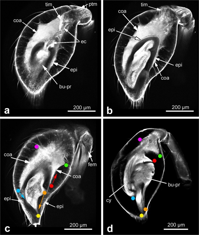
Figure 4.
Morphogenesis of the bulb primordium at mid subadult stage. (a–c) Different focal planes of the same CLSM scan to show morphological details inside of the club. The club includes two segments, tibia and tarsus. The primordium undergoes further differentiation and a structure of coagulated material is evident. (d) Club shortly before the end of the subadult stage. Important landmarks that help to understand morphogenetic movements between c and d are denoted by coloured dots. The most significant movements are indicated by coloured arrows in (c): the tip of the future cymbium rotates towards the bulb primordium (blue arrow), the ventral rim of the future haematodocha retracts proximally (red arrow), whereas the tip of the lateral protrusion (the future embolus) rotates and shifts distally to nestle to the future conductor (orange arrow). Scale bar: 200 µm in all panels. Abbreviations: bu-pr, bulb primordium; coa, coagulated material; cy, cymbium; ec, epidermal connection; epi epithelial hypodermis; fem, femoral muscles; mes, mesenchymal hypodermis; ptm, patellar muscles; ti, tibia; tim, tibial muscle.

