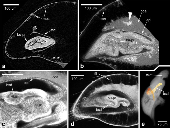
Figure 5.
Morphological details within the developing pedipalp and bulb primordium. (a) Early subadult. dice-µCT scan. Distal is to the left in all panels except panel e (proximal up). The hypodermis is epithelial near the bulb primordium, but mesenchymal along the cuticle. (b) Mid subadult. dice-µCT scan. Coagulated material contains inclusions (arrowhead) that likely represent histoblast groups. (c–e) Details of the bulb primordium at mid subadult stage, CLSM scans. (c) Detail of the base of the bulb primordium, showing the epithelial lining below the coagulated material. A part of the blind sperm duct is visible in cross section. (d) Filaments are present between the body of the coagulated material and the mesenchymal hypodermis below the cuticle. The blind sperm duct is seen in sagittal section and a portion of the epidermal connection is visible (that leads through to the tibia, not included in this focal plane). (e) 3D reconstruction of the blind sperm duct within the bulb primordium (coloured in orange). The other cavity that is visible is a portion of the epidermal connection to the tibia. Scale bars: 100 µm in a, b, d; 50 µm in c; 75 µm in e. Abbreviations: bsd, blind sperm duct; bu-pr, bulb primordium; coa, coagulated material; ec, epidermal connection; fil, filaments; epi, epithelial hypodermis; mes, mesenchymal hypodermis.

