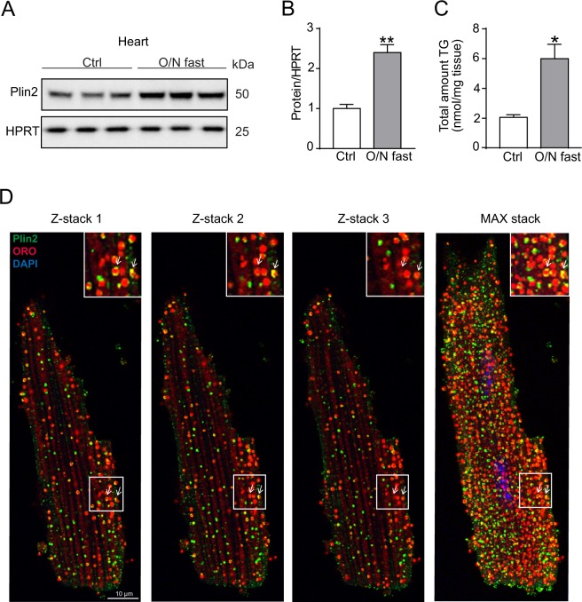
Figure 1.
Regulation of Plin2. (A) Immunoblot analysis of Plin2 using protein lysates from control (refed) and O/N fasted C57Bl/6N mice hearts (n = 3). Full-length blots are presented in Supplementary Fig. S7. (B) Quantification of immunoblot analysis on Plin2 (n = 3). (C) Triglyceride content in hearts of control (refed) and overnight fasted mice (n = 3). (D) Confocal microscopy images on C57Bl/6N cardiomyocytes, shown as z-stack 1, 2, 3 and a max stack picture containing 30 z-stacks compromised into one image. Plin2 is shown in green, ORO (LDs) in red and nuclei in blue and co-localization of LDs and Plin2 in yellow (x630 magnification, scale bar: 10 µm). Data are presented as mean ± SEM, *p < 0.05 vs. Plin2+/+, **p < 0.01 vs. Plin2+/+. TG, triglycerides; O/N, overnight.

