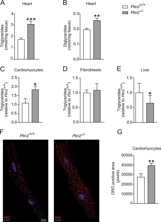
Figure 2.
Increased myocardial triglyceride accumulation in Plin2−/− hearts. (A) Triglyceride and (B) diglyceride content in hearts of 8-week-old Plin2+/+ and Plin2−/− mice after 4 hours fasting (n = 6–7). (C) Triglyceride content in cardiomyocytes isolated from Plin2+/+ and Plin2−/− hearts after O/N fast (n = 5–7). (D) Triglyceride content in fibroblasts isolated from Plin2+/+ and Plin2−/− hearts after O/N fast (n = 4). (E) Triglyceride content in liver of Plin2+/+ and Plin2−/− mice after 4 hours fasting (n = 4–5). (F) Confocal microscopy images on Plin2+/+ and Plin2−/− cardiomyocytes, shown as a merged picture containing 10 z-stacks compressed into one image. LDs are shown in red and nuclei in blue (x630 magnification, scale bar: 10 µm). (G) Quantification of ORO positive area (pixels) in Plin2+/+ and Plin2−/− cardiomyocytes (n = 4–5). Data are presented as mean ± SEM, *p < 0.05 vs. Plin2+/+, **p < 0.01 vs. Plin2+/+ and ***p < 0.001 vs. Plin2+/+; TG, triglycerides; DG, diglyceride; O/N, overnight.

